Recently, directed by academician Zhuang Songlin, professor Peng Yan from Terahertz Technology Innovation Research Institute published a paper entitled Terahertz Imaging and Spectroscopy in Cancer Diagnostics: A Technical Review in journal BME Frontiers.

In cooperation with Science Partner Journal (SPJ), BME Frontiers was released by Science/AAAS and Suzhou Institute of Biomedical Engineering and Technology, Chinese Academy of Sciences. This research is reported by official Wechat of Science.
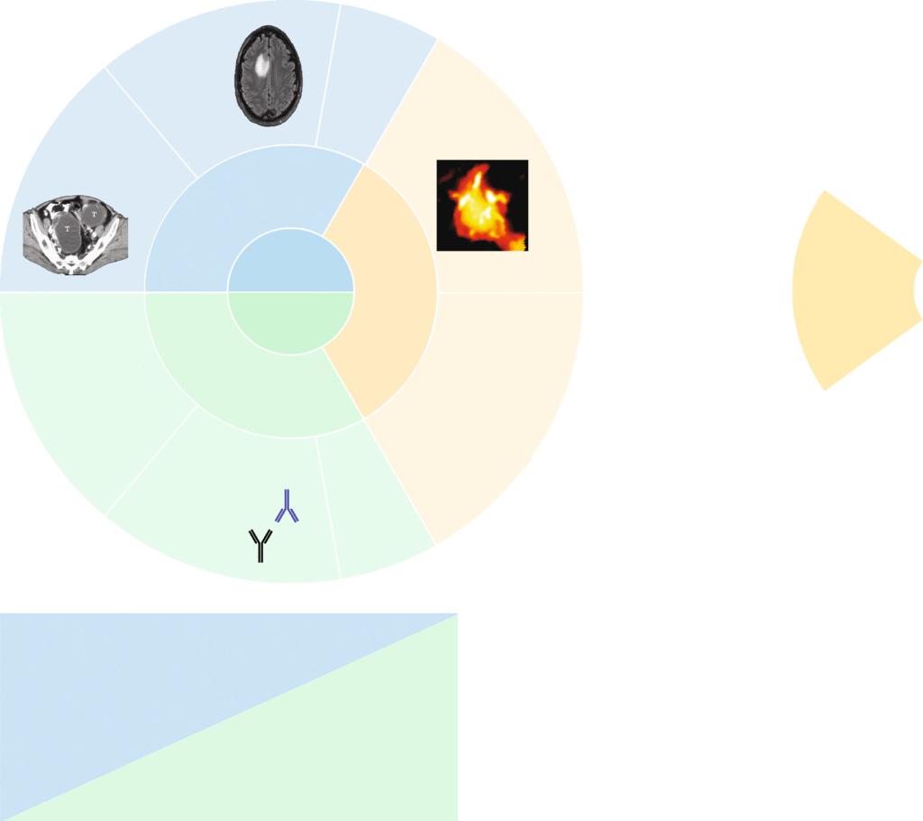
Terahertz (THz) waves are electromagnetic waves with frequency in the range from 0.1 to 10 THz [8]. As THz waves are located between the microwave and infrared regions on the electromagnetic spectrum, they exhibit features of both regions: they are nonionizing, noninvasive, and phase-sensitive to polar substances, can be used for spectral finger printing, and coherent detection, and offer good resolution (up to 50 μm) and penetration capabilities [9]. Based on these unique features, THz waves have potential applications in many fields [10–15]. In oncology, the nonionizing and noninvasive nature and good penetration capabilities of THz waves make them suitable to inspect tissue both in vivo and ex vivo with high accuracy while causing minimal harm. Furthermore, spectral fingerprinting with THz waves can enable qualitative and quantitative analysis of cancer biomarkers.

Schematic illustration of the handheld TPI probe system (from Ref. [28]).
In this paper, the project team summarizes recent studies of THz imaging and spectroscopy in cancer diagnosis from the past 5 years. They also present auxiliary methods to improve the SNR or enhance the THz signal. These works demonstrate the promise of using THz technologies for cancer diagnosis.
Because the changes in biomarker concentration intensify with the development of cancer, the quantitative analysis of biomarker concentrations can help with cancer staging. Various regression algorithms have been used to realize quantitative biomarker analysis. In 2018, Peng et al. reported a method to analyze the main substances in human brain tissue cells, including γ-aminobutyric acid, L-glutamic acid, D-myo-inositol, creatine monohydrate, cholesterol, noradrenaline, and N-acetylaspartate. When cancer occurs, the concentrations ofnoradrenaline and N-acetylaspartate will increase and decrease, respectively.

Future developments of THz technologies for cancer diagnosis include combining THz imaging with THz spectral fingerprinting of biomarkers to realize qualitative identification and quantitative analysis simultaneously. To achieve this objective, further improvements in THz systems and auxiliary methods are needed.
Link to the paper: https://spj.sciencemag.org/journals/bmef/2020/2547609
Source from School of Optical-electrical and Computer Engineering