July 12, 2021
| By Zhang Shunsheng | Copyedited by William, Zhang Liu
Optical microscopy is an important tool in the field of biology, with which researchers can obtain a variety of biological scale information in living cells and tissues. However, the diffraction limit restricts the resolution of traditional optical imaging systems, making it impossible for nanoscale (one nanometer is equal to a billionth of a meter) objects in the cells to perform optical imaging.
Stimulated Emission Loss (STED) microscopy is one of the super-resolution fluorescence technologies awarded the Nobel Prize in Chemistry in 2014. This technology can perform optical imaging for nanoscale objects, enabling researchers to carry out subcellular research. In recent years, great progress has been made in STED microscopy, but it is still a challenge to perform high-contrast, super-resolution imaging in the deep layer of biological specimens without causing light damage. In the STED microscope, organic fluorophores are often used as nanoprobes for biological samples. However, strong pulses are need to lighten them, which in turn will result in phototoxicity, photobleaching and autofluorescence. Besides, organic fluorophores usually work in the visible region, limiting STED microscope’s application in the research of deep tissues due to light attenuation and aberration.
The results of international cooperation between the University of Shanghai for Science and Technology(USST), the National University of Singapore and Jinan University have overcome these limitations and developed a new kind of luminescent lanthanide nanoprobe that can be used in super-resolution imaging for low-power STED microscopy of subcellular structures and deep tissues. This result is titled “Continuous-wave Near-IR STED Microscopy using Downshifting Lanthanide Nanoparticles”, the achievement was published in Nature Nanotechnology on June 14.
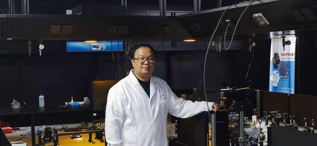
The co-first author of the paper, Professor Zhang Qiming of the University of Shanghai for Science and Technology said, “When the nanoprobe is irradiated with a second NIR laser of a different wavelength, the down conversion luminescence is almost completely exhausted, and the required beam intensity is 100 times lower than that of the organic fluorophore.” This technology can perform super-resolution optical imaging of deep tissues under the conditions of minimal phototoxicity, photobleaching, and autofluorescence. The key to realize STED in the newly developed down-conversion doped lanthanide nanoprobe is the doped neodymium emitter, which has a quasi-four-level energy configuration, as its lower excitation energy level is in the metastable state that can maintain population reversal under low-power excitation.
USST Academician Gu Min said, “These nanoprobes have the potential to expand the application range of STED microscopes. The use of low-power continuous wave irradiation can also reduce the size and cost of the imaging system, and promote the development of small portable STED microscopes in the future, hopefully enabling microscopes to play a greater role in the fields of biomedicine and super-resolution imaging.”
The research was jointly directed by Academician Gu Min from the University of Shanghai for Science and Technology, Professor Liu Xiaogang from the National University of Singapore, and Professor Li Xiangping from Jinan University. The experimental research work was jointly carried out by Dr. Liang Liangliang from the National University of Singapore, Dr. Ziwei Feng from Jinan University and Professor Qiming Zhang from the University of Shanghai for Science and Technology.